
Cancer remains one of the leading causes of death worldwide. The key determinant of prognosis isstage of tumor detection. The earlier the malignant process is diagnosed, the higher the likelihood of radical treatment, prolonged remission and preservation of the patient's quality of life.
1. Why early diagnosis is critical
The oncological process develops in stages: from precancerous changes to an invasive tumor and metastasis. Stages I–II:
tumor localized,
no or minimal metastasis,
possible organ-preserving treatment,
5-year survival is significantly higher.
Late diagnosis (stages III–IV) often requires aggressive combination therapy (surgery + chemotherapy + radiotherapy), which increases toxic load and reduces prognosis.
Oncology Principle:Early Detection = Less Intervention + Better Outcome.
2. Basic Cancer Diagnostic Techniques
Diagnosis is based on a multi-level principle: clinical examination → instrumental methods → laboratory tests → morphological verification.
2.1 Clinical Stage
Collection of anamnesis (heredity, risk factors, occupational hazards)
Physical examination
Assessment of non-specific symptoms (weakness, weight loss, anemia, pain)
2.2
Instrumental Imaging TechniquesMammography

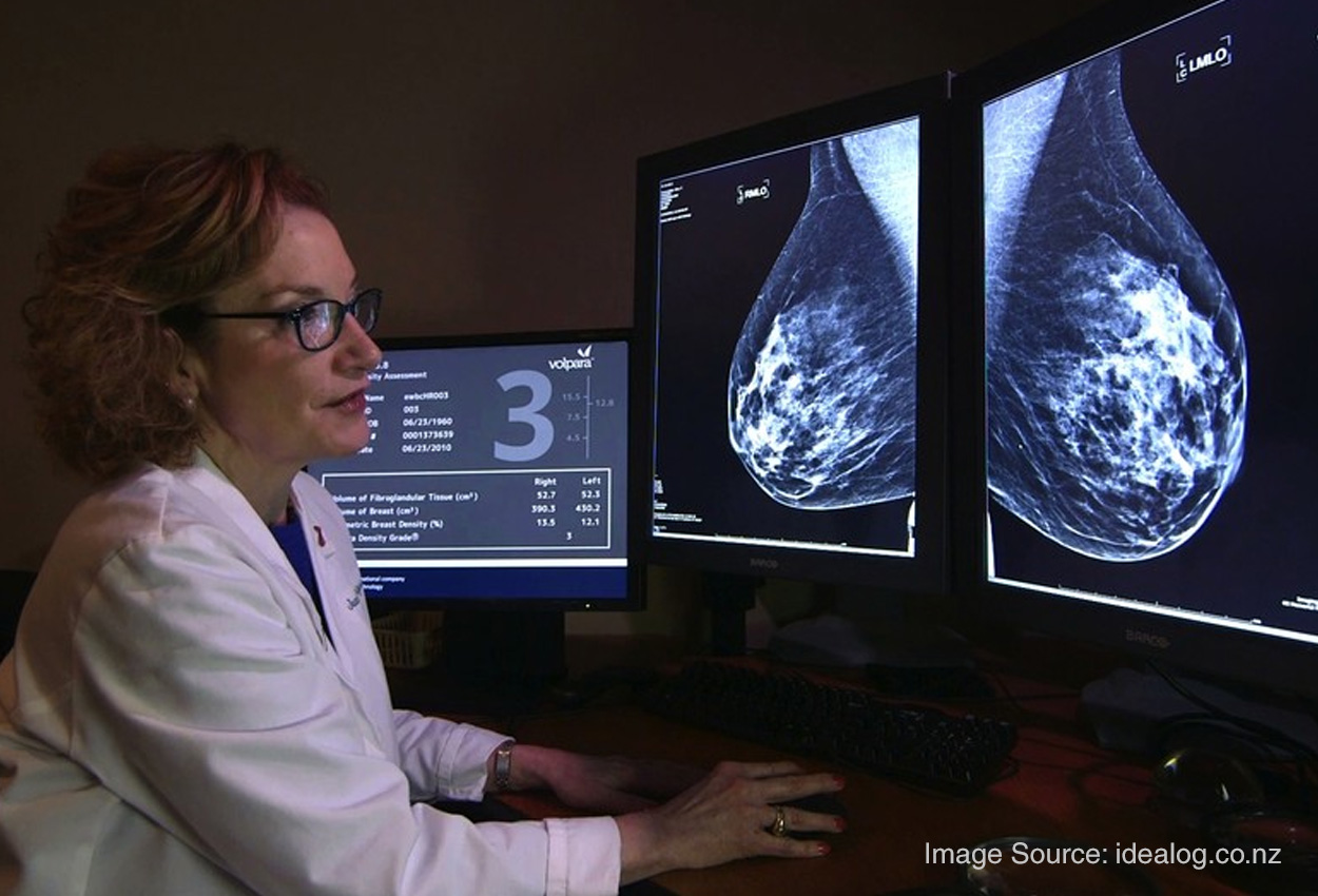

Used for early detection of breast cancer.
Allowsthe detection of microcalcifications and tumors before clinicalmanifestations.
Computed tomography (CT)


It is used to diagnose tumors of the lungs, abdominal organs, pelvis.
Low-doseCT scan is recommended for risk groups (smokers).
Magnetic Resonance Imaging (MRI)


High sensitivity in brain, pelvic, soft tissuetumors.
Endoscopic Techniques


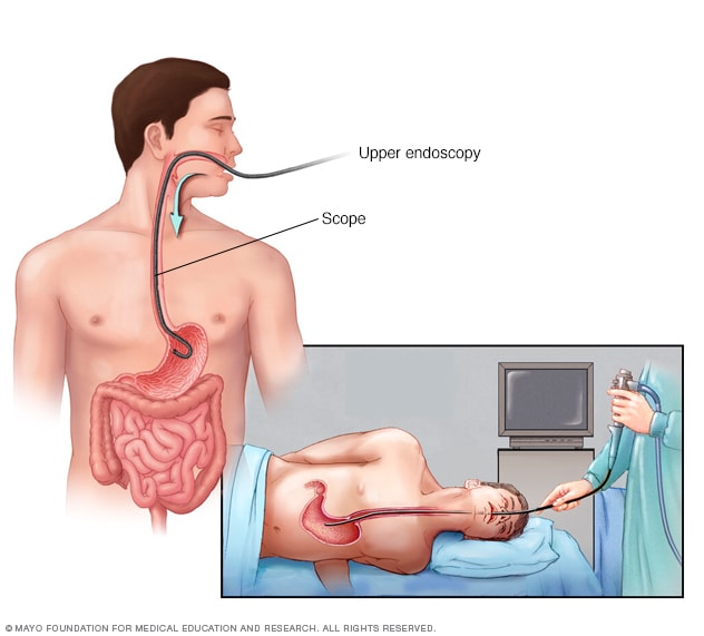
Colonoscopy — detection of intestinal cancer and polyps
Gastroscopy — diagnosis of gastric cancer
Let you perform a biopsy during theprocedure.
3.Laboratory Diagnostics
Tumor markers
Determined in blood (PSA, CA-125, CEA, etc.) .
Usedby:
for treatment monitoring,
recurrence assessments,
in combination with other .
methods
Important:tumor markers are not an independent screeningmethod.
4 Morphological Verification —
Gold StandardFinal diagnosis is made after:
Biopsy
Histological Examination
Immunohistochemistry
Molecular Genetic Analysis
Without morphological confirmation, the diagnosis of cancer is not considered established.
5.Screening is the foundation of early detection
Screening is performed in asymptomatic at-riskindividuals.
Main screening programs:
| Localization | Method | Recommended age |
|---|---|---|
| Breast cancer | Mammography | 40–70 years old |
| Cervical cancer | PAP test /HPV test | 21–65 years old |
| Colorectal cancer | Colonoscopy /occult blood test | 45+ |
| Lung Cancer | Low-dose CT | Smokers 50+ |
Regular screening reduces mortality by 20–40% depending on the type of cancer.
6.Risk Factors
Smoking
Alcohol
Obesity
Heredity
Viral infections (HPV, HBV, HCV)
Ionizing radiation
Professional carcinogens
Risk factor control is an element of primaryprevention.
7. Current Trends in Oncology Diagnostics
Molecular Diagnostics (NGS panels)
Liquid Biopsy
PET-CT for staging
Artificial Intelligence in Image Analysis
Personalized Medicine
The future of oncology is preclinical early detection and targeted therapy.
Closing
Early diagnosis of cancer is not only a medical task, but also a social responsibility. Modern technologies make it possible to detect tumors at minimal stages, when treatment is most effective.
Regular examinations, attention to symptoms, and participation in screening programs are key factors in reducing cancermortality.
Oncology is not a sentence. If detected in a timely manner, it is a manageabledisease.